
Перкутанна нефростомія (ПН) – це малоінвазивна процедура, яка виконується у спеціалізованих лікарнях. Хоча термін звучить лякаюче, сучасні методи зробили її безпечною та короткочасною — у більшості випадків перебування в лікарні становить 1–2 дні.
Якщо ви не знаєте точно, що таке нефростомія або коли її роблять, почніть звідти. Для ознайомлення прочитайте повний посібник з догляду.
Підготовка до операції
- Голодування протягом 6–8 годин перед операцією.
- Припинення прийому антикоагулянтів (Сінтром, Плавікс, НОАК) після консультації з лікарем — зазвичай за 3-5 днів до операції.
- Передопераційне обстеження: загальний аналіз крові, біохімічний аналіз, коагуляція, посів сечі.
- Профілактичні антибіотики за 30-60 хвилин до пункції.
- Отримання підписаної згоди після інформації про співвідношення ризику та користі.
Анестезія
Найпоширенішою формою є місцева анестезія у поперековій ділянці, часто поєднана з легкою внутрішньовенною седацією. У дітей, пацієнтів, які не співпрацюють, або у складних випадках використовується загальна анестезія.
При місцевій анестезії пацієнт не притомний, але не відчуває болю. Співпраця (дихання за командою) полегшує роботу інтервенційного рентгенолога.
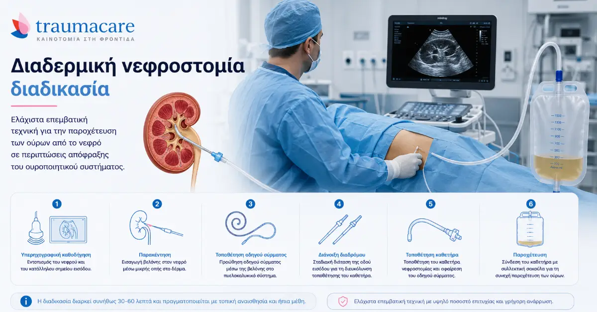
Етапи процедури
1. Положення пацієнта
Пацієнта розміщують у положенні на животі або на боці під кутом 30°. Поперекова область оголюється та обробляється антисептиком (зазвичай розчином хлоргексидину або повідоном).
2. Керівництво візуалізацією
Ідеальне місце пункції визначається за допомогою ультразвукового дослідження або флюороскопії. Метою є менший розмір балії (чашечки) на нижньому полюсі нирки — безпечніший шлях з меншим ризиком кровотечі.
3. Пункція
Використовуючи тонку голку (типу Chiba, 18 або 21 G), лікар проколює шкіру та просувається під контролем візуалізації до тазу. Правильність введення підтверджується сечею, що витікає з голки.
4. Провідник
Тонкий дріт (провідник) вводиться через голку, закріплену в тазу. Усі наступні інструменти ковзатимуть по ньому.
5. Розширення тракту
Поступово більші розширювачі використовуються для відкриття тракту від шкіри до тазу для розміщення останнього катетера.
6. Розміщення катетера
Катетер (зазвичай 8-14 Fr зі спіральним кінчиком) ковзає по дроту. У правильному положенні спіральний кінчик формується в тазу та фіксує трубку.
7. Стабілізація та підключення
Катетер фіксується швом до шкіри або за допомогою адгезивної системи StatLock. Він з'єднаний з початковим дренажним мішком. Місце виходу покривають пов'язки та прозора водонепроникна плівка.
Тривалість
Всього: 30–60 хвилин для неускладнених випадків. Більш складні випадки (пацієнти з ожирінням, незбільшена нирка, анатомічні варіації) можуть тривати довше.
Після операції
- Перебування в лікарні 24–48 годин для спостереження.
- Спочатку сеча може бути кров’янистою — це очікувано і минає протягом 24–72 годин.
- Легкий біль у попереку, який контролюється звичайними знеболювальними.
- Ультразвукове дослідження для виключення гематоми.
- Інформація пацієнта та його родини щодо догляду за сечею.
- Виписка з лікарні після прозорої сечі та підтвердження стабільності життєво важливих функцій.
Потенційні ризики
Процедура вважається безпечною, але будь-яка інвазивна процедура має ризики. Серйозні ускладнення <2% включають: значну кровотечу (1-2%), пошкодження сусідніх органів (<1%), пневмоторакс при високих проколах (<1%), сепсис (зазвичай за наявності інфекції).
Показники успіху
У збільшених нирках (гідронефроз) показники успішного розміщення перевищують 98%. У незбільшених нирках (рідкісніше показання) показники падають до 90%.
| ✉️ Контактна форма | 💬 Канал Viber Traumacare ✓ |
| Переглянути всі продукти для нефростомії → |
У вас є якісь питання? Надіслати повідомлення
Наша команда відповість вам протягом 24 годин. Ми є ексклюзивними представниками B Braun Avitum та дистриб'ютором Convatec, з контрактом EOPYY з 2014 року.
Контактна форма
Джерела: Грецьке товариство інтервенційної радіології, Загальна лікарня Патри, Урядовий вісник B' 5395/2025. Інформаційна стаття, не замінює медичної консультації.